


Explore Scientific Smart Microscope Slide: Dragonfly Wing Stained Specimen (English)
| English | Français | Deutsche | Nederlandse | Italiano | Polskimi | Portuguesas | Español | 中国 | 日本人 | 한국어 |

First appearing on earth about 300 million years ago, Dragonflies are carnivorous insects. Adults have large, multifaceted eyes, a long body, and two pairs of strong, transparent wings, sometimes with colored patches. And it's the wings that have scientists excited.

A recent discovery about the nature of the structure of the Dragonfly's wing has been discovered under very high-powered microscopes, showing nanostructures that destroy bacteria. Scientists hope that they can learn from these structures so that they can make similar surfaces for the next generation smart materials that can also destroy bacteria, especially for use in medical applications to prevent infections in patients.
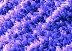
False color scanning helium-ion microscope image of more than 10 billion tiny nanostructures on a dragonfly wing.Image credit: Annalena Wolff
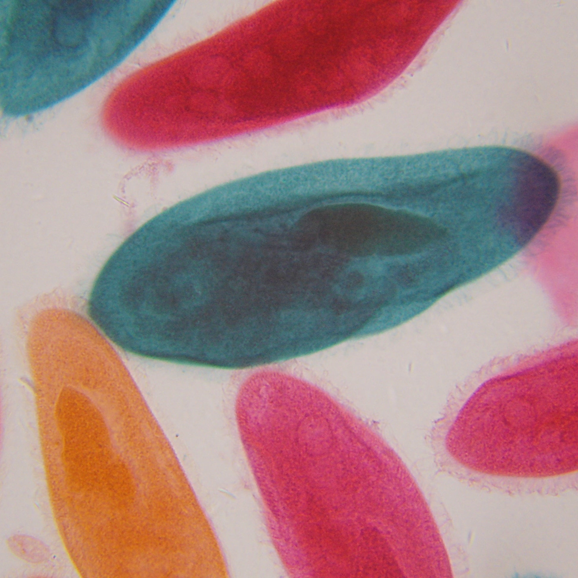
While only scanning helium-ion microscopes can show these nanostructures, it is fascinating to examine the intricate structure of the wing. The aqua blue color in this specimen is from Methelyne Blue stain used to highlight the structure against the background. Specimen staining is just one technique to help students researching microscopic specimens to determine what they are (their morphology).

See all Microscopes from Explore Scientific